



In the realm of regional anesthesia and interventional diagnostics, the spinal needle is a masterclass in micro-precision manufacturing. Performing a subarachnoid block or diagnostic lumbar puncture requires penetrating dense ligaments, traversing the epidural space, and puncturing the delicate dural-arachnoid membrane to access cerebrospinal fluid (CSF)—all without causing vascular laceration, nerve root injury, or debilitating persistent fluid leaks.
For medical device procurement managers, OEM/ODM engineers, and clinical directors, understanding the structural and fluid dynamics of spinal needles is not merely an academic exercise. Needle tip geometry, metallurgical yield strength, lumen fluid dynamics, and stylet tolerances directly dictate clinical outcomes, most notably the incidence of Post-Dural Puncture Headache (PDPH).
This guide delivers an exhaustive technical breakdown of spinal needle classifications, tip mechanics, gauge parameters, international regulatory standards, and manufacturing best practices.
1. The Anatomy of Dural Puncture and Clinical Fundamentals
Neuraxial Mechanics and Fluid Dynamics
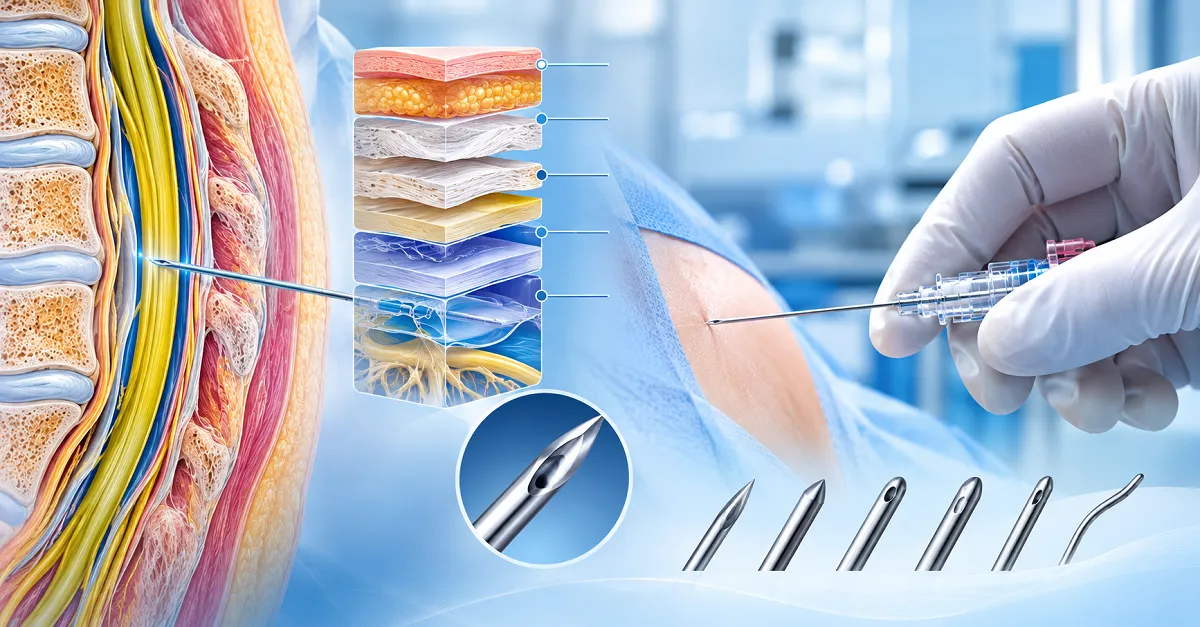
A spinal injection requires transversing seven distinct anatomical layers: skin, subcutaneous tissue, supraspinous ligament, interspinous ligament, ligamentum flavum, epidural space, and the dural-arachnoid sac.
Skin ➔ Subcutaneous Tissue ➔ Supraspinous Ligament ➔ Interspinous Ligament ➔ Ligamentum Flavum ➔ Epidural Space ➔ Dura-Arachnoid Maternal ComplexThe human dura mater is an anisotropic fibro-elastic membrane measuring between 0.3 mm and 0.5 mm in thickness at the lumbar levels (L3–L5). Contrary to traditional anatomical beliefs that dural collagen fibers run purely longitudinally, modern scanning electron microscopy demonstrates that the dura comprises concentric lamellae of collagen and elastic fibers arranged in multidirectional, interwoven patterns.
When a needle used for lumbar puncture or spinal anesthesia breaches this multi-layered matrix, fluid dynamics govern the outcome:
- Hydrostatic Drainage: Normal CSF opening pressure ranges from 10 to 18 cm $H_2O$ in lateral recumbency. When a puncture site fails to seal immediately, CSF weeps into the epidural space at a rate exceeding production (~0.35 mL/min or ~500 mL/day).
- Cerebral Traction: The depletion of the CSF fluid cushion causes downward displacement (traction) of pain-sensitive intracranial structures—specifically the sensory meninges, tentorium cerebelli, and cerebral bridging veins—when the patient assumes an upright posture.
- Cerebral Vasodilation: According to the Monro-Kellie hypothesis, intracranial blood volume increases secondarily to compensate for lost CSF volume, provoking bifrontal or bioccipital orthostatic cephalalgia (PDPH).
Clinical Objectives: Flashback vs. Trauma
Anesthesiologists face a dual challenge: maximizing the speed of CSF visualization in the needle hub (which requires a wide inner diameter) while minimizing dural trauma and persistent leakage (which demands the narrowest possible outer profile and an atraumatic profile).
2. Core Classification: Cutting (Traumatic) vs. Non-Cutting (Atraumatic) Tips
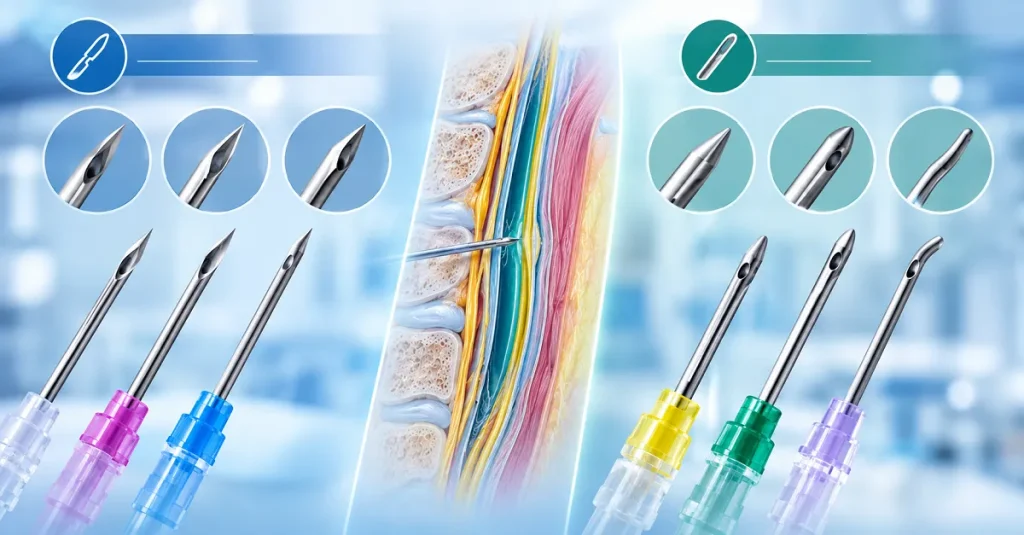
The primary taxonomy of modern spinal needles divides instruments by tip architecture: cutting (traumatic) ve non-cutting / pencil-point (atraumatic) designs.
A. Cutting Spinal Needles (Sharp / Beveled Geometry)
Cutting needles feature an open, beveled end with ground cutting edges designed to slice cleanly through tissue planes with minimal axial penetration force.
1. The Quincke Spinal Needle
The Quincke profile is the oldest and most widely recognized geometry.
- Tip Architecture: Features a medium-angle, sharp, three-facet beveled lancet tip. The ground cutting facets meet at an acute point, with an inner stylet ground flat to match the bevel’s plane precisely.
- Insertion Dynamics: Slices through fibrous connective tissue and the dura mater like a scalpel. Because it encounters low tissue resistance, it provides tactile awareness of anatomical strata, though with less distinct “dural pop” contrast than blunt conical profiles.
- Clinical Pros: Excellent directional tracking; low resistance in calcified interspinous ligaments (frequently encountered in geriatric patients); exceptional utility in emergency diagnostic taps (Quinke needle for lumbar puncture).
- Clinical Cons: Leaves an open, clean-cut defect in the dural membrane that heals slowly and permits sustained CSF weeping. If inserted with the bevel perpendicular to longitudinal tissue fibers, it severs fibers rather than parting them, escalating PDPH rates to 10%–30% in high-risk populations.
2. The Greene Spinal Needle
- Tip Architecture: Developed as an intermediate design, the Greene needle features a curved, rounded non-cutting bevel contour with a lateral aperture profile.
- Insertion Dynamics: Designed to mitigate dural slicing by blunting the lateral edges of the bevel while preserving a sharp distal point.
- Clinical Performance: Lower PDPH incidence than a standard Quincke, but displaced in modern practice by true pencil-point options due to higher manufacturing complexity without matching pencil-point safety metrics.
3. Spinocan Needles
- Design Specifics: B. Braun’s proprietary variation of the Quincke needle. The Spinocan spinal needle features an optimized micro-ground lancet bevel angle and ultra-smooth surface finish designed to reduce initial puncture force while maximizing needle stability and stylet flushness.
B. Non-Cutting / Pencil Point Needles (Atraumatic Geometry)
Pencil-point designs eliminate cutting edges entirely at the puncture point. The distal end terminates in a solid, rounded cone, and fluid transfer occurs through an electrochemically polished side hole (lateral eye) machined proximal to the conical apex.
1. The Whitacre Spinal Needle
- Tip Architecture: Features a rigid conical tip tapering symmetrically to a rounded, non-cutting apex. The lateral discharge eye is located approximately 1 to 2 mm proximal to the needle tip.
- Insertion Dynamics: Spreads and parts the collagen and elastic fibers of the dura mater rather than transecting them. Upon needle withdrawal, the parted elastic dural fibers recoil inward, rapidly occluding the puncture channel.
- PDPH Mitigation: The clinical gold standard for obstetrics and ambulatory surgery. A 25g whitacre spinal needle reduces the incidence of PDPH to below 1.5%–2%.
- Tactile Feedback: Produces a distinct “snap” or “pop” as the blunt conical tip overcomes the tension of the ligamentum flavum and breaches the dura.
2. Pencan Needles
- Design Specifics: Manufactured by B. Braun, the pencan spinal needle features a precision-engineered conical tip with an optimized tip-to-hole distance. The geometry provides high structural rigidity to resist tip deflection (skiving) and curling during ligamentous transit, paired with rapid CSF detection.
3. Sprotte Needles
- Design Specifics: Pioneered by Pajunk (Pajunk spinal needle), the Sprotte features an elongated, smooth lateral eye with rounded internal borders. This maximises cross-sectional flow area, accelerating CSF flashback even through ultra-fine 27G lumens.
C. Anatomy-Specific Hybrid: The Tuohy Needle
Bu Tuohy spinal needle (more accurately classified as an epidural needle) features a hollow, curved distal tip (Schenker/Tuohy curve).
- Mekanizma: The directional curve deflects the sharp tip away from the dura, allowing tactile localization of the epidural space via loss-of-resistance (LOR) techniques and facilitating the tangential passage of an epidural catheter.
- Combined Spinal-Epidural (CSE): Modern regional anesthesia sets frequently utilize a long, low-gauge pencil-point needle threaded directly through the lumen of a Tuohy needle (needle-through-needle technique).
Tip Mechanical Comparison
| Technical & Clinical Metric | Quincke Needle (Cutting) | Whitacre / Pencan (Pencil Point) | Sprotte (Elongated Lateral Eye) |
| Distal Geometry | Sharp, tri-beveled cutting lancet | Solid symmetrical cone; side eye | Long ogival conical tip; large side eye |
| Dural Action | Cuts / severs dural lamellae | Dilates and separates fibers | Separates fibers atraumatically |
| Insertion Force Required | Low (0.8−1.2 N) | Moderate (1.8−2.8 N) | Moderate (1.5−2.2 N) |
| Tactile “Dural Pop” | Subtle to absent | Crisp, pronounced | Distinct, clear |
| PDPH Rate (25G–27G) | 5.0%−12.0% | 0.5%−2.0% | 0.5%−1.8% |
| Tissue Coring Propensity | Moderate (if stylet misaligned) | Negligible | Negligible |
| Deflection Resistance | Moderate (bevel induces drift) | High (symmetrical cone tracks straight) | Moderate to High |
3. Needle Gauge (G), Dimensions, and ISO Color Coding
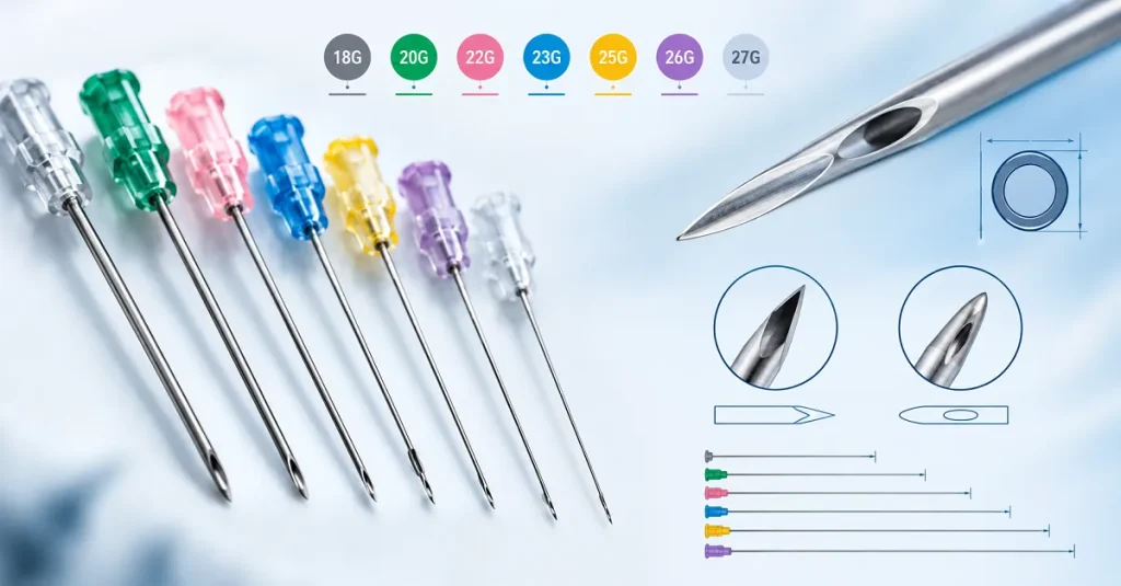
Reducing the cannula from a 22G to a 27G outer diameter reduces tissue trauma, but the smaller inner lumen drastically slows CSF flashback. Precision manufacturing must maintain an ultra-thin wall (UTW) configuration—thinning the cannula wall without compromising metallurgical collapse pressure or column buckling strength.
Detailed Size, Gauge, and Color-Code Matrix
Medical-grade spinal needles adhere to ISO 6009 for hub and packaging color identification.
| Gösterge (G) | Color Code (ISO 6009) | Nominal OD (mm) | Tolerance OD (mm) | Recommended Application | Typical Insertion Technique |
| 18G | Pembe | 1.2 | ±0.02 | Specialized drainage, epidural, fluid aspiration | Direct insertion without guide |
| 20G | Sarı | 0.9 | ±0.015 | High-viscosity CSF, neurosurgery taps | Direct insertion |
| 21G | Koyu Yeşil | 0.8 | ±0.015 | Emergency diagnostic tap, therapeutic drainage | Direct insertion |
| 22G | Siyah | 0.7 | ±0.01 | Diagnostic LP, geriatric spinal block | Direct insertion |
| 24G | Mor | 0.55 | ±0.01 | Standard regional spinal anesthesia | Optional introducer |
| 25G | Turuncu | 0.5 | ±0.01 | Obstetric, orthopedic, daylight anesthesia | Introducer recommended |
| 26G | Kahverengi | 0.45 | ±0.008 | Low-trauma subarachnoid block | Introducer required |
| 27G | Medium Grey | 0.4 | ±0.008 | Ultra-low PDPH target (pediatric/young adult) | Introducer mandatory |
Cannula Lengths and Bariatric Challenges
The nominal adult standard is the 3.5-inch spinal needle (90 mm). However, global demographic trends and rising obesity rates demand a wider portfolio of operational lengths:
- Pediatric / Neonatal: 25 mm to 50 mm (1 to 2 inches).
- Standard Adult: 90 mm (3.5 inches).
- Extended / Bariatric: 120 mm to 150 mm (4.75 to 6 inches).
The Introducer Spinal Needle
When utilizing ultra-fine atraumatic cannulas (25G, 26G, and 27G), the needle lacks the structural rigidity needed to puncture tough keratinized skin, subcutaneous adipose tissue, and fibrotic supraspinous ligaments without bending.
Bir introducer spinal needle (typically 20G or 22G, measuring 32 mm to 38 mm in length) resolves this limitation:
- The rigid, beveled introducer punctures the skin and anchors into the interspinous ligament.
- The long, fine-gauge spinal needle is then inserted coaxially through the introducer’s lumen.
- The introducer provides lateral stability and structural tracking, preventing cannula deflection (skiving) and ensuring that the atraumatic tip meets the dura perpendicularly.
4. International Brand Benchmarks vs. OEM Engineering
Global regional anesthesia is anchored by several established brands. OEM and contract medical device manufacturers analyze these market references to engineer equivalent or superior products.
Competitive Manufacturing Matrix
       B. Braun (Spinocan / Pencan)        Pajunk (Sprotte Innovations)
    ┌────────────────────────────────┐  ┌────────────────────────────────┐
    │ High-precision grinding hubs   │  │ Pioneering atraumatic eye      │
    │ Prism/Facet visualization tech │  │ Polished inner eye contours    │
    │ German-drawn cannula tubing    │  │ Integrated echo-reflectors     │
    └────────────────────────────────┘  └────────────────────────────────┘
                                  ▲    ▲
                                  │    │
                     OEM Contract Engineering
    ┌────────────────────────────────────────────────────────────────────┐
    │ • Equivalent 304/316L tensile yield strength                       │
    │ • Custom Luer lock transparency & zero-dead-space chambers         │
    │ • Scalable ISO 13485 cleanroom automated manufacturing             │
    └────────────────────────────────────────────────────────────────────┘
                                  │    │
                                  ▼    ▼
        Nipro / Romsons Supplies           Egemen / Specialist Producers
    ┌────────────────────────────────┐  ┌────────────────────────────────┐
    │ Scalable global logistics      │  │ Specialized neuraxial trays    │
    │ Competitive cost per unit      │  │ Custom length / gauge options  │
    │ Robust hospital standard lines │  │ Regional kit integration       │
    └────────────────────────────────┘  └────────────────────────────────┘
- B. Braun Spinal Needles: Renowned for the Spinocan (cutting) and Pencan (pencil-point). Their benchmark feature is the ergonomic hub with a clear view chamber that provides magnified, early CSF visualization through faceted prism geometry.
- Pajunk Spinal Needles: Celebrated for the Sprotte atraumatic geometry. Pajunk utilizes specialized cold-forming and electrochemical polishing to ensure the side-eye margins are completely smooth, preventing tissue coring and dural snagging.
- Nipro Spinal Needle & Romsons Spinal Needle: The high-volume workhorses of global healthcare systems. These manufacturers emphasize high automated throughput, uniform siliconization, and competitive pricing for tender contracts.
- Egemen Spinal Needle: Known for regional anesthesia convenience packs and needle systems tailored for precision anesthetic delivery across emerging and established clinical centers.

5. Engineering, Metallurgy, and Cleanroom Manufacturing
To match the precision required for neuraxial access, spinal needle manufacturing demands strict compliance with international medical standards, advanced metallurgy, and zero-defect quality control.
   Raw Coil Stock (AISI 304/316L)
                 │
                 ▼
     Tube Drawing & Sizing (ISO 9626)
                 │
                 ▼
       Multi-Axis CNC Grinding (Electrochemical Deburring)
                 │
                 ▼
   Stylet-to-Cannula Zero-Tolerance Fitting
                 │
                 ▼
    Solvent-Free Ultrasonic Cleaning & Micro-Siliconization
                 │
                 ▼
   Hub Molding (PC/PP) & Adhesive Curing (ISO 80369-7)
                 │
                 ▼
 Class 100,000 Cleanroom Packaging & EtO Sterilization (ISO 11135)
Metallurgical Foundations: ISO 9626 Cannula Tubing
Needle cannulas are drawn from cold-worked, seamless austenitic stainless steel:
- AISI 304: High tensile strength (1800 – 2200 MPa), providing the column strength needed to resist axial buckling under insertion load.
- AISI 316L: Lower carbon and added molybdenum (2%–3%) offer superior pitting resistance and ductility, reducing the risk of cannula micro-cracking during deep ligament transit.
Multi-Axis Grinding and Stylet Alignment
- Burr-Free Micro-Machining: Bevels and side eyes are ground using high-speed diamond-impregnated vitrified wheels or electrochemical machining (ECM). This eliminates micro-burrs, backward metal hooks, and rough internal kerfs that could shred dural fibers or biopsy epidermal tissues.
- Zero-Step Stylet Fit: A critical quality indicator in a premium disposable spinal needle is the mechanical flushness between the stylet and the outer cannula tip. Any step-off creates a cutting shelf that cores tissue, increasing the risk of introducing an epidermal plug into the subarachnoid space (which can lead to iatrogenic spinal epidermoid tumors).
Fluid Flow and Hub Architecture (ISO 80369-7)
Needle hubs are injection molded from medical-grade, lipid-resistant transparent polycarbonate (PC) or polypropylene (PP).
- Early Flashback Detection: The hub’s internal geometry incorporates a conical, low-dead-space chamber that channels the first micro-drop of CSF into view.
- ISO 80369-7 Compliance: Connectors utilize 6% Luer lock tapers designed to prevent cross-connections with non-vascular or enteral lines, ensuring leak-free coupling with standard syringes and manometers.
Siliconization and Force Profile Testing
Needles undergo micro-aerosolization of medical-grade polydimethylsiloxane (silicone oil) complying with ISO 7864. This low-friction coating reduces insertion drag through tough ligaments by up to 40%, enhancing patient comfort and tactile sensitivity for the clinician.
Penetration Force (N) Profile Across Anatomical Strata
   Force (N)
     │
 3.0 ┼                       /\ (Ligamentum Flavum)
     │                      /  \
 2.0 ┼      /\ (Skin)      /    \
     │     /  \           /      \       /\ (Dural "Pop")
 1.0 ┼    /    \_________/        \_____/  \_________ Subarachnoid Space
     │   /                                           (Flashback!)
 0.0 ┴──────────────────────────────────────────────────────────► Depth
Sterility and Infection Control
- Paketleme: Needles are sealed in individual medical-grade paper/film pouches (Tyvek/PET) that preserve a sterile barrier under variable humidity and pressure.
- Sterilizasyon Doğrulaması: Validated to achieve a Sterility Assurance Level (SAL) of 10-6 using Ethylene Oxide gas (ISO 11135) or Gamma irradiation (ISO 11137).
- Safety Integration: Modern hospitals increasingly adopt safety spinal needles, which feature spring-loaded or sliding cannula locks to permanently encase contaminated sharp tips post-procedure, eliminating needlestick injuries (NSI).

Sıkça Sorulan Sorular (SSS)
The Quincke needle features a sharp, tri-beveled cutting lancet tip that slices through tissue and dural fibers, requiring lower insertion force but leaving an open dural defect. The Whitacre needle features a solid, non-cutting conical (pencil-point) tip with a lateral discharge aperture. It parts dural fibers atraumatically rather than severing them, substantially reducing CSF leakage and lowering the risk of Post-Dural Puncture Headache (PDPH).
Fine-gauge needles have thin outer diameters (0.40 mm–0.50 mm) and high flexibility. Without support, they tend to bend or deflect (skive) when encountering skin and fibrous interspinous ligaments. A rigid 20G or 22G introducer needle penetrates the superficial tissue to provide a stable, straight guide channel, ensuring the fine spinal needle reaches the subarachnoid space without bending.
Needles measuring 25G to 27G—particularly when combined with pencil-point profiles (Whitacre, Pencan, Sprotte)—provide the lowest clinical rates of PDPH, consistently documented below 1%–2%. By contrast, larger cutting needles (such as a 20G or 22G Quincke) carry PDPH risks ranging from 10% to over 30%, especially in younger and obstetric populations.
Tissue coring occurs when a hollow needle shears off a cylinder of skin, fat, or ligamentous tissue during insertion, which can then be carried into the subarachnoid space. Manufacturers prevent coring by fitting a precision-ground, flush inner stylet that matches the outer bevel angle with zero tolerance, eliminating open cutting edges along the inner circumference during entry.
Spinal needle hubs are molded from medical-grade transparent polycarbonate (PC) or clarified polypropylene (PP) to provide clear, immediate visualization of cerebrospinal fluid (CSF) flashback. Rapid flashback confirms subarachnoid entry before injecting local anesthetics or collecting fluid samples.
A Tuohy needle is primarily designed for epidural anesthesia. It features a curved (directional) tip engineered to identify the epidural space via loss of resistance and guide an epidural catheter tangentially without puncturing the dura. While not used alone for primary spinal injection due to its large gauge (16G–18G), it is commonly used in Combined Spinal-Epidural (CSE) procedures as the outer guide conduit for a longer, fine-gauge spinal needle.
Spinal needle hubs follow ISO 6009 (Hypodermic needles for single use — Color coding for identification). This standard assigns specific colors to nominal outer diameters (e.g., 18G is Pink, 22G is Black, 25G is Orange, and 27G is Medium Grey), helping clinicians immediately verify the needle gauge.
AISI 304 stainless steel offers higher tensile strength and structural hardness, providing excellent column stability to resist buckling during insertion. AISI 316L contains lower carbon and adds 2%–3% molybdenum, which increases ductility and enhances corrosion resistance against harsh pharmaceutical solutions, though it exhibits slightly lower tensile yield strength.
Bizi ziyaret edin:https://topkeymedical.com/
E-posta:info@topkeymedical.com