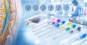



In the manufacture of tıbbi iğneler, clinical performance is governed at the sub-millimeter scale. While macroscopic specifications—such as nominal gauge, outer diameter (OD), inner diameter (ID), and usable cannula length—dominate procurement catalogs, the micro-geometry of the needle tip dictates the physical interface between medical device and biological substrate. Specifically, the needle bevel angle, facet configuration, and heel treatment govern insertion mechanics, tissue shearing dynamics, penetration force profiles, and particulate generation.
For medical device engineers, original equipment manufacturers (OEMs), clinical educators, and procurement directors, understanding needle tip mechanics is critical. Whether performing a perpendicular intramuscular depot injection, cannulating a fragile pediatric vein, or accessing a chlorobutyl rubber vial stopper, the interplay between the needle’s manufactured bevel grind and the clinician’s procedural insertion angle directly influences patient trauma, tissue displacement, hematoma formation, and foreign-body particulate contamination.
1. The Micro-Mechanics of Needle Tip Geometry
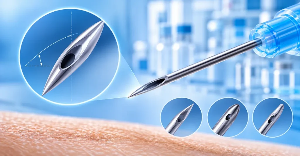
A hypodermic needle tip is not a simple angled cylinder. It is a compound three-dimensional profile generated through multi-axis precision grinding.
TOP VIEW (LANCET / TRI-BEVEL)
---------------------------------------------
                 /|\
 Secondary      / | \   Primary    <-- Primary Bevel Face
 Lancet Facets /  |  \  Bevel
              /   |   \
-------------+----+----+---------------------
                  ^
             Needle Apex

SIDE VIEW (LONGITUDINAL PROFILE)
        Primary Grind Angle (Theta)
            \       /
             \     /
              \   /
               \ /
----------------\----------------------------
                 \              Outer Cannula Wall
------------------\
                   \   Lumen
--------------------\
         ^           \          Inner Cannula Wall
    Cutting Heel      \----------------------
                       ^
                  Needle Point
Primary, Secondary, and Tertiary Grinds
- Primary Bevel Face: The initial angled plane generated by slicing the drawn stainless steel cannula at a predetermined angle relative to the tube’s longitudinal axis.
- Secondary Lancet Facets: Two lateral grinds made by rotating the cannula symmetrically along its central axis and applying secondary abrasive cuts. These lateral facets converge at the distal apex to create a lancet point.
- The Cutting Heel (Heel of the Bevel): The proximal boundary where the ground bevel terminates and transitions back into the cylindrical inner lumen and outer circumference of the tubing. If left untreated, this junction presents a razor-sharp, crescent-shaped cutting edge.
Mechanics of Penetration: Slicing vs. Tearing
When a solid or hollow instrument breaches biological tissue, the required mechanical work is defined by the penetration force applied over the displacement distance:
Work (W) = Integral of Force F(x) over Displacement Distance dx
The physical force profile during injection consists of three distinct mechanical phases:
- Pre-puncture Tent-like Deformation: The distal tip contacts the skin (stratum corneum) or vascular wall, stretching the tissue elastically until local shear stress exceeds ultimate tensile yield strength.
- Puncture and Crack Initiation: The apex of the needle breaches the surface, initiating an axial crack.
- Cannula Transit and Frictional Drag: The widening bevel facets expand the tissue aperture while the outer cannula shaft slides through the incision, encountering frictional resistance.
A finely honed, multi-faceted bevel with optimized primary and secondary angles functions as a true micro-scalpel. It incises tissue cleanly along cellular planes. Conversely, a blunt, unoptimized, or poorly deburred bevel acts as an unsharpened wedge: it stretches, tears, and compresses cellular bundles, triggering localized release of inflammatory cytokines, activating nociceptive pain pathways, and leaving a ragged, slow-healing wound tract.
2. Understanding Bevel Grind Types & Engineering Classifications
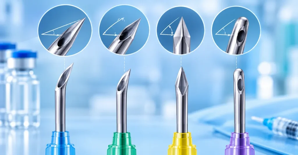
Standard hypodermic needles are categorized into distinct structural classes governed by international standards such as ISO 7864 (Sterile hypodermic needles for single use) and ISO 9626 (Stainless steel needle tubing for the manufacture of medical devices).
Needle Bevel Profiles:
- Regular Bevel (A-Bevel): 10 to 12 degrees angle | Long profile | Low entry force | IM / SC / Autoinjectors
- Short Bevel (B-Bevel): 18 to 20 degrees angle | Short profile | Reduced depth | Nerve blocks / ABG
- Blunt / Anti-Coring Bevel: 40 to 45 degrees angle | Deflected heel | Vial transfer | Zero coring
A. Regular Bevel (A-Bevel / Medium Bevel)
- Geometry: The primary bevel angle ranges from 10 to 12 degrees (measured against the long axis of the cannula), producing an elongated, slender elliptical tip profile.
- Mechanical Characteristics: The acute entry angle minimizes initial peak penetration force. By extending the cutting length along a greater longitudinal distance, tissue layers are separated gradually.
- Primary Applications: Standard intramuscular (IM) and deep subcutaneous (SC) hypodermic injections, high-volume vaccination campaigns, and pre-filled syringe (PFS) autoinjector platforms.
- Engineering Trade-offs: While regular bevels require lower puncture force, the tip is structurally thinner. When encountering dense cortical bone or tough fascial planes, an unsupported 10-degree tip is susceptible to tip curling (“fish-hooking”).
B. Short Bevel (B-Bevel / Low-Angle Bevel)
- Geometry: The primary angle is ground more steeply, typically between 18 and 20 degrees, producing a compact, stubby bevel profile.
- Mechanical Characteristics: The distance from the distal tip to the fully open lumen (bevel length) is substantially shorter than that of an A-bevel.
- Primary Applications: Regional anesthesia, peripheral nerve blocks, arterial blood gas (ABG) sampling, and specialized epidural placements.
- Clinical Justification: In close proximity to neural structures or vessel back-walls, an elongated 10-degree bevel can simultaneously reside in two distinct anatomical compartments (e.g., partially inside a vessel lumen and partially in the extravascular space). A short bevel ensures that the entire fluid orifice enters the target structure immediately upon tactile breach, preventing back-wall transfixion (vessel perforation).
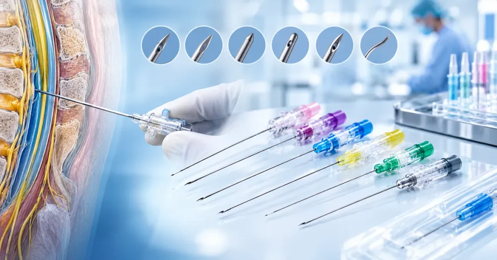
C. Tri-Beveled / Lancet Point
- Geometry: A compound geometry combining a primary grind with two symmetric secondary side facets ground at angles between 15 and 25 degrees relative to the vertical plane.
- Mechanical Characteristics: Instead of presenting a single elliptical plane, the cutting edge is segmented into razor-sharp cutting facets that meet at a central point.
- Clinical Impact: Modern phlebotomy and vascular access devices rely almost universally on tri-beveled grinds. Studies demonstrate that a true tri-beveled tip reduces peak skin penetration force by 30% to 50% compared to a basic single-facet grind, substantially mitigating procedural pain.
D. Blunt Fill & Anti-Coring Bevels
- Geometry: Machined at steep angles (40 to 45 degrees) or featuring custom deflected/non-coring heel treatments.
- Function: Designed exclusively for fluid aspiration, vial reconstitution, and medication compounding. These needles lack the sharpness required to puncture human skin, protecting healthcare workers from needlestick injuries (NSIs) while preventing the mechanical punching of rubber stoppers.
3. The Coring Effect: Causes, Risks, and Preventive Engineering
The “coring effect” is one of the most persistent technical challenges in pharmaceutical compounding and clinical needle design.
THE CORING MECHANISM ON ELASTOMERIC VIAL CLOSURES
Step 1: Unfavorable Angle / Sharp Heel
Cannula Axis () enters rubber stopper.
Sharp heel acts like a punch.
Step 2: Shearing and Plug Formation
As the sharp trailing heel enters, it shears a cylindrical
rubber plug into the liquid solution, forming a dangerous particulate embolus.
Pathomechanics of Coring
When a hollow cannula enters an elastomeric closure (such as a rubber vial stopper) or biological tissue perpendicularly without proper technique or tip optimization, the open lumen acts as a circular punch:
- The distal point pierces the elastomer, elastically displacing the rubber laterally.
- As the needle advances, the elastomeric material rebounds into the expanding open bevel lumen.
- When the sharp trailing edge—the cutting heel—passes through the plane of the closure, it acts as a die cutter. It shears off a cylindrical fragment of rubber or tissue matching the inner diameter of the cannula.
Clinical and Pharmaceutical Consequences
- Vial Coring & Micro-Emboli: When reconstituted medication is drawn into a syringe alongside microscopic rubber particles, intravenous administration can cause pulmonary micro-embolisms, systemic vascular occlusions, granulomatous tissue reactions, and chemical phlebitis. The United States Pharmacopeia (USP 788 Particulate Matter in Injections) and ISO 8871-5 impose rigorous thresholds on permissible particulate matter in parenterals.
- Tissue Coring & Epidermoid Implantation: In regional anesthesia and lumbar punctures, coring tissue during skin transit can transport epidermal plugs into the subarachnoid or epidural space. These viable epithelial cells can engraft and proliferate, forming iatrogenic spinal epidermoid tumors years after the procedure.
Engineering Solutions to Coring
Medical needle manufacturers deploy several metallurgical and geometric strategies to eliminate coring:
- Heel Blunting: Precision micro-blasting or laser ablation is applied specifically to the proximal heel of the bevel. This dulls the circular cutting edge without affecting the primary and secondary cutting facets at the distal tip.
- Deflected / Huber Points: The distal point is curved slightly inward toward the cannula’s central axis (as seen in Huber needles used for implanted ports). The solid heel deflects elastomeric material laterally rather than slicing through it.
- Reverse Back-Beveling: Grinding a micro-relief on the trailing heel to eliminate the sharp shearing corner.
- Electrochemical Deburring: Dissolves micro-feathered wire edges along the heel perimeter during post-grinding processing.
- Optimized Lubrication (Siliconization): Applying an ultra-thin layer of medical-grade polydimethylsiloxane minimizes friction-induced deformation of elastomeric closures, reducing the shear stress that triggers coring.
4. Clinical Insertion Angles: Correlating Bevel Position with Procedural Success
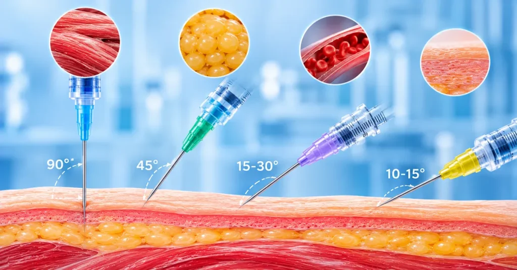
A needle’s manufactured bevel works in tandem with the clinician’s procedural insertion angle. The angle of attack dictates how tissue lamellae are loaded, divided, or preserved.
INJECTION ANGLE HIERARCHY

90 Degrees  --> Intramuscular (IM) / Deep Target
45 Degrees  --> Subcutaneous (SC) / Adipose Layer
15 to 30 Degrees --> Phlebotomy / Venipuncture
10 to 15 Degrees --> Intradermal (ID) / Fragile Winged Butterfly
A. Intramuscular (IM) Injections: The Perpendicular Approach
- Clinical Standard: Delivered at a 90-degree angle relative to the skin surface (90 degree needle injection).
- Anatomical Target: Deep vascularized skeletal muscle beds (deltoid, ventrogluteal, vastus lateralis) bypassing the superficial epidermis, dermis, and subcutaneous adipose tissue.
- Mechanical Rationale: A perpendicular vector represents the shortest direct linear path to deep muscle beds. Entering at 90 degrees ensures that the entire usable length of a 1-inch or 1.5-inch cannula is utilized. A 90-degree approach with an elongated 10 to 12-degree regular bevel parts the longitudinally aligned muscle fibers with minimal transverse muscle tearing, promoting the formation of an encapsulated medication depot and preventing subcutaneous back-tracking.
B. Subcutaneous (SC) Injections: Managing Adipose Depth
- Clinical Standard: Delivered at a 45-degree angle trajectory, or at 90 degrees when using specialized ultra-short micro-needles (4 mm to 8 mm).
- Anatomical Target: The loose, vascular connective tissue of the hypodermis/adipose layer beneath the dermis.
- Mechanical Rationale: Standard 5/8-inch (16 mm) hypodermic needles inserted perpendicularly in cachectic, pediatric, or lean adult patients carry a high risk of accidentally penetrating underlying muscle tissue. Injecting at 45 degrees alters the effective depth:
Effective Depth = Cannula Length multiplied by Sine of Insertion Angle
For a 16 mm needle inserted at 45 degrees: Effective Depth = 16 mm multiplied by 0.707 = approximately 11.3 mm
This ensures consistent placement within the subcutaneous layer, guaranteeing predictable, sustained pharmacokinetic drug absorption.
C. Phlebotomy and Blood Draw: Vessel Stabilization and Bevel-Up Protocol
- Clinical Standard: Executed at a 15 to 30-degree phlebotomy needle angle with the bevel facing upward (Bevel-Up).
- Anatomical Target: Superficial veins of the antecubital fossa (median cubital, cephalic, basilic veins).
- Hemodynamic and Vascular Mechanics:
- Bevel-Up Entry: Orienting the bevel upward allows the razor-sharp distal point to breach the anterior elastic vascular wall first. The opening of the lumen enters the bloodstream smoothly, establishing prompt blood aspiration.
- Preventing Vessel Transfixion: Inserting the needle at an angle steeper than 30 degrees significantly increases the likelihood of punching entirely through the posterior wall of the vessel (“blowing the vein”), causing immediate blood extravasation, severe pain, and hematoma formation.
- Angle Adjustment for Shallow Veins: For superficial, fragile veins, the entry angle must be flattened to 15 to 20 degrees to ensure the entire bevel resides within the vascular lumen without scraping the inner endothelial layer.
PHLEBOTOMY: BEVEL-UP INSERTION MECHANICS (15 to 30 Degrees)

Skin Surface: ----------------------------------------------
Cannula Axis:       \ (15 to 30 Degrees)
                     \
                      \  Bevel UP [Lumen Open to Blood Flow]
=======================\===================== Anterior Vein Wall
                        \ <-- Lancet Tip Enters Lumen Smoothly
                         \
                          No Transfixion!
============================================= Posterior Vein Wall
D. Peripheral IV Cannulation and Butterfly Infusion Sets
- Clinical Standard: The IV needle angle is typically initiated at 10 to 25 degrees, while the butterfly needle angle is maintained at an ultra-low 10 to 15 degrees.
- Clinical Mechanics: Winged infusion (butterfly) needles and peripheral intravenous catheters (PIVC) are deployed in delicate peripheral vasculature (e.g., dorsal venous network of the hand, pediatric scalp veins, geriatric sclerotic vessels).
- The Transition Step: Upon observing early blood flashback in the transparent visualization chamber, the clinician drops the cannula almost parallel to the skin surface (5 to 10 degrees) and advances the instrument an additional 1 mm to 2 mm. This ensures that both the needle point and the catheter tip clear the vessel wall before the flexible cannula is threaded forward over the needle.
5. Comprehensive Clinical and Engineering Reference Matrix
| Clinical Procedure / Application | Manufactured Bevel Type | Primary Grind Angle (θ) | Clinical Insertion Angle | Standart Gösterge Aralığı | ISO 6009 Hub Color Code | Primary Mechanical & Clinical Objective |
| Intramuscular (IM) Injection | Regular Bevel (A-Bevel) | 10°–12° | 90∘ | 21G–23G | Green / Black / Blue | Deep muscular penetration; minimizes tracking; low tissue tearing |
| Subcutaneous (SC) Injection | Regular / Tri-Bevel | 10°–12° | 45∘ (or 90∘ if ≤8 mm) | 25G–27G | Orange / Medium Grey | Targets adipose layer; avoids intramuscular vascular absorption |
| Phlebotomy / Venipuncture | Tri-Bevel Lancet Point | 11°–14° | 15∘–30∘ (Bevel-Up) | 21G–22G | Green / Black | Clean vascular entry; prevents posterior wall transfixion; minimizes pain |
| Butterfly Infusion Set | Short Bevel / Tri-Bevel | 14°–17° | 10∘–15∘ | 23G–25G | Deep Blue / Orange | Accesses fragile, small-caliber veins; low insertion profile |
| Intravenous (IV) Catheter | Back-Cut / V-Point Bevel | 12°–15° | 10∘–25∘ (Drop to 5∘) | 16G–24G | Grey / Green / Pink / Blue / Yellow | Smooth catheter transition; instant flashback; avoids blowing the vein |
| Intradermal (ID) Injection | Short Bevel (B-Bevel) | 18°–20° | 5∘–15∘ (Bevel-Up) | 26G–27G | Brown / Medium Grey | Forms localized dermal wheal (bleb); avoids subcutaneous transit |
| Arterial Blood Gas (ABG) | Short Bevel (B-Bevel) | 18°–20° | 45∘–60∘ (Radial) | 22G–25G | Black / Blue / Orange | Punctures high-pressure arterial wall; avoids shearing artery back-wall |
| Vial Reconstitution / Transfer | Blunt Fill / Anti-Coring | 40°–45° | 45∘ initial, advance to 90∘ | 18G | Red / Pink / Purple | Pierces rubber stopper without shearing; eliminates particulate coring |
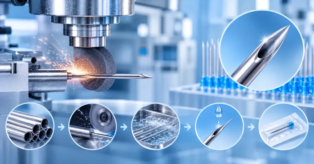
6. Manufacturing Precision: CNC Grinding, Deburring, and Siliconization
Engineering medical needles that reliably achieve low tissue trauma and zero coring requires tightly controlled, multi-stage manufacturing systems.
   Raw Drawn Stainless Tubing (ISO 9626)
                    │
                    ▼
   Electrolytic Cut-Off & Deburring
                    │
                    ▼
   Multi-Axis CNC Grinding (Facet Geometry: ±0.5° Tolerance)
                    │
                    ▼
   Electrochemical Polishing (Removes Micro-Burrs & Wire Edges)
                    │
                    ▼
   Heel Blunting & Micro-Ablation Treatment
                    │
                    ▼
   Ultrasonic Solvent Degreasing & Cleaning
                    │
                    ▼
   Micro-Aerosolized Siliconization (ISO 7864 Compliant)
                    │
                    ▼
   Automated 100% Machine-Vision Optical Inspection
High-Speed Multi-Axis CNC Grinding
Needle cannulas are secured in high-density fixture blocks and advanced through multi-station abrasive grinding wheels:
- Abrasive Wheels: Vitrified bond diamond or cubic boron nitride (CBN) wheels running at high surface speeds.
- Angular Control: High-precision multi-axis CNC machines maintain bevel angle tolerances within +/- 0.5 degrees, ensuring uniform tip geometry across production volumes of millions of units.
- Coolant Management: Synthetic flood coolants prevent localized metallurgical overheating, phase changes, and thermal softening of cold-worked 304 or 316L stainless steel.
Electrochemical Polishing and Deburring
Mechanical grinding leaves microscopic metal remnants known as “feathered burrs” or “wire edges” along the ground facets and inside the open lumen heel. If not removed, these burrs tear tissue during insertion and snag during needle withdrawal.
- Electrochemical Machining (ECM): The ground cannulas are subjected to an electrolytic acid bath where electrical current dissolves micro-burrs preferentially from exposed high-energy edges. This yields an exceptionally smooth, mirror-like surface finish with a roughness average (Ra) of less than 0.1 micrometers.
The Role of Micro-Layer Siliconization
Uncoated stainless steel displays a surprisingly high coefficient of friction when contacting biological tissues and elastomeric polymers.
- Coating Specification: Manufacturers apply medical-grade polydimethylsiloxane (silicone oil) meeting ISO 7864 and USP Class VI standards via micro-aerosolization or ultrasonic dipping.
- Force Reduction: A validated silicone coating forms a durable, biocompatible molecular layer that reduces dynamic drag and withdrawal resistance by up to 50%. This preserves structural tissue alignment and prevents the needle from pulling or distorting the dermal matrix upon retraction.
7. Sourcing and Customization Guide for OEM/ODM Buyers
When sourcing medical needles or partnering with contract manufacturers for custom cannula development, procurement teams should evaluate suppliers across four core engineering dimensions:
1. Cannula Tubing Dimensions and Wall Architecture (ISO 9626)
- Regular Wall (RW): Standard structural thickness; provides maximum column strength and buckling resistance for long needles or dense tissue penetration.
- Thin Wall (TW) and Extra-Thin Wall (ETW): Cannula outer diameter remains constant while inner diameter is expanded. This allows for higher volumetric flow rates or permits the use of a smaller outer gauge without compromising injection speed.
- Ultra-Thin Wall (UTW): Demands high-tensile, high-purity medical-grade austenitic stainless steel (AISI 304 or 316L) to prevent lumen collapse under high injection pressures (e.g., automated contrast injection or high-viscosity biologics).
2. Custom Bevel Engineering and Application Matching
- Aesthetics and Mesotherapy: Demands ultra-fine gauges (30G, 32G, 34G) ground with custom tri-bevel profiles to minimize micro-trauma, facial bruising, and procedural downtime.
- Ophthalmology: Requires specialized micro-bevels ground to exact angular tolerances to prevent elevated intraocular pressure or corneal shearing during sub-Tenon and intravitreal delivery.
- Autoinjectors and Pre-Filled Syringes (PFS): Demand rigid staking of cannulas into glass or cyclic olefin polymer (COP) barrels with consistent bevel orientation relative to syringe graduations.
3. Comprehensive Quality Assurance and Inspection Standards
A reliable OEM partner must provide verified documentation and continuous automated quality control, including:
- Automated Machine Vision Inspection: High-speed inline cameras capable of 360-degree inspection for tip deformities, inverted hooks, dimensional tolerances, and incomplete deburring.
- Particulate Monitoring: Lot-by-lot testing for sub-visible particulate matter complying with USP 788 and USP 787.
- Coring Benchmarks: Standardized testing verifying that puncture of rubber closures complies with ISO 8871-5 (zero visible elastomeric cores observed across sequential vial penetrations).
- Penetration Force Testing: Continuous load-cell testing measuring peak puncture and dynamic sliding force through calibrated polyurethane foil substrates simulating human skin per ISO 7864.
Sıkça Sorulan Sorular (SSS)
The needle bevel angle is a permanent physical parameter ground into the stainless steel cannula during manufacturing (such as an acute 10 to 12-degree regular bevel or an 18 to 20-degree short bevel). The clinical insertion angle is the dynamic angle at which a healthcare provider introduces the needle relative to the patient’s skin surface or a vial closure (such as entering at 90 degrees for an intramuscular injection or 15 to 30 degrees for phlebotomy).
A tri-beveled needle incorporates three distinct precision-ground facets: a primary bevel face and two secondary lateral cutting facets. When entering tissue, these facets function as microscopic cutting blades that slice cleanly through tissue planes with minimal axial resistance. A single-facet needle tends to stretch and tear tissue laterally, requiring higher penetration force and triggering a stronger response from pain receptors (nociceptors) in the dermis.
Coring occurs when the hollow inner cutting heel of a needle shears out a cylindrical core of rubber from the vial stopper. This usually happens when a sharp needle is inserted perpendicularly (90 degrees) without prior deflection. It can be prevented clinically by using the “bevel-up” technique at a 45-degree angle until the heel passes through the stopper before rotating to 90 degrees, or industrially by using engineered anti-coring needles with blunted, electrochemically deburred heels or 40 to 45-degree blunt fill designs.
Short bevels feature a compact tip profile where the lumen opening is contained within a very short longitudinal distance. In nerve blocks, this provides crisp tactile feedback when crossing fascial planes while reducing the risk of penetrating or lacerating the nerve perineurium. In arterial blood gas (ABG) sampling, a short bevel ensures the entire open lumen sits within the narrow arterial lumen, avoiding simultaneous puncture of the posterior vessel wall.
Inserting a needle with the bevel facing up ensures that the sharp distal point punctures the upper vascular wall first, allowing the opening of the lumen to enter the blood vessel smoothly. If inserted bevel-down, the point tends to scrape along the anterior endothelial wall, requiring deeper penetration to achieve blood return. This increases the risk of transfixing the vessel (“blowing the vein”) and causing painful hematomas.
No. Standard intramuscular (IM) injections must be delivered at a 90-degree angle relative to the skin. Inserting the needle at 45 degrees limits penetration depth, increasing the risk that the medication will be deposited into the subcutaneous adipose layer instead of deep muscle tissue. This can cause poor drug absorption, localized sterile abscesses, tissue necrosis, or reduced vaccine efficacy.
Sterile hypodermic needles are governed by ISO 7864 (Sterile hypodermic needles for single use — Requirements and test methods). This standard outlines stringent requirements for stainless steel tubing, dimensional tolerances, allowable lubricant/silicone quantities, limit thresholds for particulate matter, freedom from burrs and hooks, and standardized test methods for measuring penetration force through calibrated substrates.
Applying a microscopic layer of medical-grade polydimethylsiloxane (silicone oil) reduces the coefficient of friction between the stainless steel cannula and tissue or elastomeric stoppers. A properly siliconized needle reduces peak penetration force and dynamic drag resistance by up to 50%. This lowers pain on entry and withdrawal, prevents tissue from adhering to the cannula, and minimizes the shear forces that generate tissue and rubber coring.